
Table of Contents
Scientists Confirm a Hidden Protein Helps Tumors Survive Chemotherapy
For generations, the foundational strategy behind cancer treatments like chemotherapy and radiation has been straightforward and brutal: inflict massive, catastrophic damage to the DNA inside malignant cells. When the genetic blueprints inside a tumor cell are shattered beyond recovery, the cell loses its ability to replicate and naturally self-destructs. For many patients, this is the primary line of defense against aggressive malignancies.
However, oncologists have long been frustrated by a baffling phenomenon: some cancer cells manage to absorb heavy hits from treatment, patch up their wounds, and continue dividing, leading to dangerous treatment resistance.
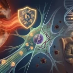
A groundbreaking study published in the medical journal Genes & Development has uncovered a major reason why this defensive shield exists. Researchers have discovered that a notorious protein called MYC—long infamous for driving rapid tumor growth—leads a secret double life. When chemotherapy shatters a cancer cell’s genetic code, this protein rushes directly to the fracture site to orchestrate an emergency cellular cleanup, helping the tumor survive the very treatments designed to destroy it. This discovery sheds new light on treatment resistance, particularly for incredibly challenging conditions like pancreatic cancer.

Scientists Confirm a Hidden Protein Helps Tumors Survive Chemotherapy
The Double Life of the MYC Protein
To understand why this discovery is reshaping how scientists look at tumor biology, it helps to look at how MYC operates under normal circumstances. Mechanistically, MYC functions as a transcription factor (a specialized protein that acts as a master switchboard to control which genes are turned on or off inside a cell).
[ Traditional View of MYC ] ───> Controls Gene Expression ───> Drives Rapid Growth & Fuel Burning
[ New Discovery ] ─────────────> Travels to DNA Breaks ───────> Recruits Repair Proteins (BRCA1, RAD51)
In a healthy body, MYC keeps cellular replication running smoothly. In a cancerous state, however, this protein becomes hyperactive. It forces the malignant cell to grow, split, and consume cellular fuel at a frantic, chaotic pace.
Yet, researchers have historically grappled with a confusing paradox. The sheer speed of growth driven by MYC naturally creates massive internal stress and structural DNA damage inside the tumor. Logically, this self-inflicted stress should cause the cancer cell to collapse. Senior study author Rosalie Sears from Oregon Health & Science University, alongside first author Gabriel Cohn, currently at the University of Würzburg, focused heavily on this contradiction.
Their research revealed that MYC doesn’t just sit safely inside the cell’s genetic control center. Instead, a modified variation of the protein actively migrates to physical breaks in the DNA strands to call for reinforcements. It functions much like a local arsonist who simultaneously keeps the local fire department on speed dial, protecting the tumor from its own chaotic growth and external medical interventions.
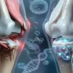
The Anatomy of a Genetic Fracture
DNA is the ultimate instruction manual for everything a cell does. Under normal conditions, minor scratches or single-strand tears to this blueprint occur constantly and are easily mended by standard cellular maintenance.

The real danger arises when a cell suffers a double-strand break—an injury where both sides of the ladder-like genetic structure snap in the exact same location. Without an intact opposite strand to serve as a template, the cell struggles to figure out how to patch the missing data.
When a tumor cell encounters extensive double-strand breaks from an IV chemotherapy drip, its internal repair bill becomes too high to pay. If the cell cannot mend these breaks, it dies.
However, if a tumor can deploy an emergency repair crew quickly enough, it can bridge these genetic gaps before the self-destruct sequence is triggered. This rapid patching is exactly how a tumor builds resistance to therapy, allowing mutated cells to survive the initial chemical onslaught and continue multiplying throughout the body.
The Chemical Security Badge: What is pS62-MYC?
The researchers discovered that MYC’s ability to transition from a growth driver to a genetic welder depends entirely on a biological process called phosphorylation (the addition of a tiny chemical tag to a protein that completely changes its behavior).
In this specific mechanism, an enzyme attaches a phosphate tag to one precise location on the MYC protein, creating a variation known as pS62-MYC. This tiny modification acts like a high-level security badge, allowing the protein to enter structural emergency zones that are normally off-limits.
Once the tagged pS62-MYC arrives at a double-strand DNA break, it directly coordinates with two well-known cellular repair proteins:
BRCA1: A prominent tumor suppressor protein involved in recognizing and organizing responses to major DNA damage.
RAD51: An enzyme that physically assists in knitting broken strands of genetic material back together.
The study demonstrated that cancer cells carrying high concentrations of this chemically tagged MYC variant were significantly more efficient at repairing shattered DNA. This structural efficiency allowed them to comfortably survive extreme environmental stress and aggressive chemotherapy exposures that would easily wipe out standard cells.
Why This Matters for Pancreatic Cancer Patients
While this cellular escape route can occur across various tumor types, the findings are exceptionally vital for the fight against pancreatic cancer. According to data tracked by the Surveillance, Epidemiology, and End Results (SEER) program, the five-year relative survival rate for pancreatic cancer in the United States sits at a sobering 13.7%.
| Patient Profile (Pancreatic Cancer) | Current 5-Year Survival Rate (SEER Data) | Primary Clinical Hurdles |
| All Combined Stages | 13.7% | • Late-stage initial detection • Dense protective tumor tissue • Intense resistance to standard chemotherapy |
Pancreatic cancer stands out as one of the most notoriously difficult conditions to treat because it is frequently diagnosed long after it has begun to spread, and its cells are inherently resistant to standard frontline treatments.
As lead author Gabriel Cohn points out, MYC activity is exceptionally high within pancreatic tumors. This discovery doesn’t imply that MYC is responsible for every single failed treatment outcome. However, it proves that the protein hands malignant cells a powerful survival tool at the exact micro-second that medical therapies are trying to destroy them.
Exposing a New Target for Future Cancer Drugs
For over thirty years, the MYC protein has deeply frustrated pharmaceutical developers. Because it has a smooth, shifting physical structure, it lacks the distinct “docking pockets” that traditional small-molecule medications use to lock onto a target. Furthermore, because healthy cells rely on basic MYC functions to survive, completely shutting down the protein nationwide would cause toxic, life-threatening side effects for the patient.
This new discovery completely shifts the strategy. Instead of attempting to eliminate the MYC protein entirely, researchers can now focus on blocking the specific repair-related pS62-MYC variant or disrupting the emergency pathway it builds with BRCA1 and RAD51. This represents a highly precise, elegant approach to drug development.
[ Classic Approach ] ───> Total MYC Elimination ───> High Toxicity & Damage to Healthy Cells
[ Modern Target ] ──────> Block pS62-MYC Repair ───> Disarms Tumor Defense Without Widespread Harm
Early progress is already underway. A 2024 phase 1 clinical trial published in Nature Medicine evaluated an innovative MYC inhibitor called OMO-103 in 22 patients living with advanced solid tumors, reporting promising early signs of drug stabilization and safety.
Building on that momentum, an active, early-phase clinical trial is currently comparing tumor biopsies from patients with advanced pancreatic ductal adenocarcinoma before and after direct exposure to OMO-103. This trial aims to see if the drug can successfully disarm the tumor’s internal repair system.
Conclusion
This newly mapped escape route doesn’t mean that traditional oncology tools like chemotherapy, radiation, or surgery are obsolete. Instead, it provides global researchers with an accurate map of a tumor’s secret underground bunker. In the complex world of oncology, having a clearer map makes all the difference. The next step is determining whether blocking this genetic repair helper can make standard chemotherapy significantly more effective, allowing doctors to hit tumors harder while sparing patients from unnecessary systemic harm.
Frequently Asked Questions
Does this discovery mean current chemotherapy treatments are ineffective?
No, chemotherapy remains an incredibly powerful and essential tool in modern cancer care. Chemotherapy successfully destroys billions of cancer cells by breaking their DNA structure. This study simply identifies a specific molecular backup mechanism that a percentage of smart tumor cells use to survive. Understanding this mechanism allows scientists to create companion drugs that block the escape route, making traditional chemotherapy far more effective.
What is the difference between a normal protein and a transcription factor like MYC?
While many proteins serve as structural building blocks or simple metabolic enzymes, a transcription factor functions as a high-level manager within the cell nucleus. It binds directly to specific segments of DNA to dictate which genetic instructions are read and transcribed into action. Because transcription factors control large networks of cellular behavior, when one goes rogue—like MYC does in cancer—it can trigger widespread cellular chaos.
Are the BRCA1 proteins mentioned in this study the same as the “breast cancer genes”?
Yes, they are the exact same proteins. The BRCA1 gene normally acts as a protective tumor suppressor that helps repair damaged DNA to prevent cancer from forming. However, inside an active tumor, the hyperactive MYC protein essentially hijacked this natural repair machinery (BRCA1 and RAD51), turning a protective corporate asset into a shield that helps the cancer cell survive chemotherapy.
How long does it typically take for a laboratory discovery like this to become an available treatment?
Transitioning a fundamental laboratory discovery into an approved, widely available clinical therapy typically spans several years. The process requires extensive pre-clinical testing, followed by three distinct phases of human clinical trials to carefully verify safety, ideal dosages, and real-world efficacy. However, because MYC inhibitors like OMO-103 are already being evaluated in active early-phase human trials, the timeline for this specific field is moving much faster than average.
Can diet or lifestyle modifications lower high MYC protein activity in the body?
No. Hyperactive MYC expression within a tumor is driven by deep somatic genetic mutations and structural chromosomal re-arrangements inside the cancer cells themselves. It cannot be regulated, lowered, or altered through dietary changes, lifestyle shifts, or over-the-counter herbal supplements. Managing altered protein pathways requires targeted, scientifically engineered pharmaceutical therapies designed to work at the molecular level.