Table of Contents
- 1. The Science of Radiomics: Seeing the Invisible
- 2. Outperforming Human Specialists in Blind Clinical Trials
- 3. Why a 16-Month Lead Time is a Lifesaving Window
- 4. Current Technical Limitations and the Path to Clinical Adoption
- 4.1. Managing False Positives
- 4.2. Next Steps: The AI-PACED Clinical Trial
- 5. Conclusion: Turning Everyday Data into Tomorrow’s Shield
- 6. Frequently Asked Questions
- 6.1. Is the REDMOD AI tool currently available at my local hospital?
- 6.2. Does this mean everyone should get a CT scan to screen for pancreatic cancer?
- 6.3. What is the difference between a normal tumor scan and a radiomics scan?
- 6.4. Why does a sudden diabetes diagnosis relate to pancreatic cancer?
- 6.5. How does the AI help if there is still no definitive cure for pancreatic cancer?
AI Identifies Early Signs of Pancreatic Cancer Years Before Diagnosis
Pancreatic cancer is one of the most formidable challenges in modern oncology. Because the pancreas sits deep within the abdomen, the disease is notoriously difficult to detect in its infancy, frequently growing silently without causing noticeable symptoms. By the time a patient experiences warning signs like jaundice, abdominal pain, or unexplained weight loss, the malignancy has often already advanced or metastasized to distant organs, severely limiting effective treatment interventions.
According to estimates from the National Institute of Cancer’s SEER program, approximately 67,530 individuals in the United States will be diagnosed with pancreatic cancer in 2026, and 52,740 will succumb to the disease. Strikingly, only about 15 percent of cases are caught while the cancer is still localized to the pancreas. Over half of all diagnoses occur after the disease has already reached a distant stage.
However, a groundbreaking study published in the medical journal Gut reveals that an advanced artificial intelligence model could fundamentally shift this diagnostic timeline. Developed by researchers at the Mayo Clinic and the University of Texas MD Anderson Cancer Center, the AI system can detect subtle, sub-visual anomalies hidden within standard abdominal CT scans—flagging future cancer cases up to three years before a human physician can spot a tumor.

AI Identifies Early Signs of Pancreatic Cancer Years Before Diagnosis
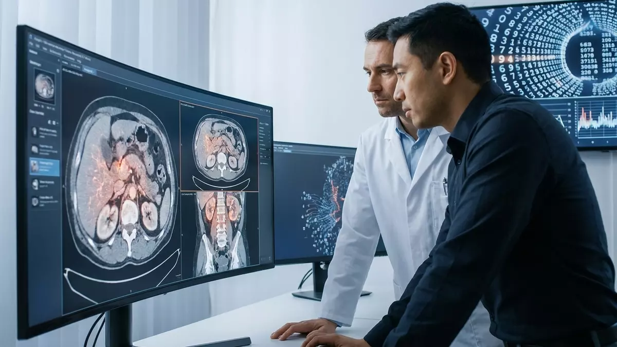
The Science of Radiomics: Seeing the Invisible
The newly developed tool is known as REDMOD, an acronym for Radiomics-based Early Detection Model. Rather than functioning like a standard computer vision tool that simply searches for a defined mass or a visible lump, REDMOD utilizes an advanced field of medical data analysis called radiomics.
Radiomics converts standard, cross-sectional medical images into high-dimensional data pipelines. The AI algorithm evaluates microscopic variations in tissue density, complex surface textures, and cellular spatial relationships that are entirely imperceptible to the human eye.

Instead of looking for a fully formed tumor, REDMOD detects the biological “footprints in the snow”—the minute, structural shifts in pancreatic tissue health that occur as healthy cells first begin to transform into a malignant state.
“The greatest barrier to saving lives from pancreatic cancer has been our inability to see the disease when it is still curable,” notes senior author Dr. Ajit Goenka, a radiologist and nuclear medicine specialist involved in the study. REDMOD directly addresses this barrier by redefining what constitutes an early warning sign.
Outperforming Human Specialists in Blind Clinical Trials
To rigorously train and validate the machine learning model, researchers supplied REDMOD with an initial training set of 969 pancreatic CT scans from individuals who eventually developed cancer, alongside healthy controls.

To ensure the AI’s efficacy extended beyond its own training data, the team conducted a strict independent blind test on an entirely separate cohort of images. This validation group consisted of 63 scans from patients who were later clinically diagnosed with pancreatic adenocarcinoma, paired with 430 control scans from individuals who remained entirely cancer-free.
Crucially, every single one of these historical scans had been previously cleared as completely normal and healthy by human radiologists at the time of the original scan.
| Diagnostic Reviewer | Detection Efficacy Rate on “Normal” Scans |
| Independent Radiologist Review | 38.9% of future cancer cases identified |
| REDMOD AI System | 73.0% of future cancer cases identified |
REDMOD correctly flagged 46 of the 63 future cancer cases, achieving an impressive 73 percent detection rate. When two expert radiologists re-examined the exact same images for the study, they were only able to retrospectively identify early irregularities in 38.9 percent of the cases. The artificial intelligence performed at nearly double the rate of human specialists.
Why a 16-Month Lead Time is a Lifesaving Window
The study revealed that REDMOD identified these microscopic tissue anomalies an average of 16 months before a clinical diagnosis was officially made. In several instances, the AI successfully flagged the silent warnings more than two years prior, with data suggesting the model can reliably predict structural pancreatic deviations up to three years in advance.
This expanded timeline could completely rewrite a patient’s prognosis. Data compiled by the American Cancer Society underscores the critical nature of early detection:
Pancreatic Cancer 5-Year Relative Survival Rates:
- Caught at the Localized Stage: 44% Survival Rate
- Caught at the Distant/Metastatic Stage: 3% Survival Rate
If a patient undergoes a routine abdominal CT scan for an unrelated health issue—such as investigating general gallbladder issues, kidney stones, or persistent stomach discomfort—REDMOD could seamlessly analyze the existing data in the background. By alerting physicians to microscopic cellular shifts long before a physical tumor forms, patients gain a vital window to undergo proactive monitoring, molecular testing, or early surgical intervention while the disease is still highly treatable.
Current Technical Limitations and the Path to Clinical Adoption
Despite its revolutionary potential, the developers of REDMOD emphasize that the system is not yet ready to serve as a widespread, commercial screening tool for the general public.
Managing False Positives
During the independent validation trial, REDMOD flagged 81 of the 430 healthy control scans as suspicious. In a real-world medical setting, these false positives could trigger unnecessary psychological distress for patients, alongside a cascade of expensive follow-up imaging, blood panels, and invasive biopsies. Balancing the algorithm’s sensitivity to minimize these false alarms is a primary focus for the research team.
Next Steps: The AI-PACED Clinical Trial
To validate the model’s safety and clinical utility moving forward, researchers are launching a comprehensive prospective study called AI-PACED. Published reports from the BMJ Group indicate that this next phase will actively test the algorithm in live healthcare settings, specifically targeting high-risk patient demographics. This includes older adults presenting with sudden, unexplained weight loss or individuals navigating a late-in-life diagnosis of type 2 diabetes—both of which are documented metabolic precursors to pancreatic malignancies.
Conclusion: Turning Everyday Data into Tomorrow’s Shield
The development of REDMOD represents a fundamental paradigm shift in how medical science approaches aggressive oncology. Rather than waiting for a silent disease to manifest as a symptomatic physical mass, healthcare is moving toward a predictive model that uncovers microscopic vulnerability hidden within the medical records and imaging files doctors already collect.
While further prospective trials are mandatory before REDMOD becomes a staple in diagnostic imaging centers, this technology marks a massive leap forward, transforming routine medical snapshots into powerful, proactive shields for patient health.
Frequently Asked Questions
Is the REDMOD AI tool currently available at my local hospital?
No, REDMOD is currently in the advanced research and clinical trial phase and is not yet approved for standard commercial use in hospitals or imaging centers. It must first undergo prospective validation in real-world healthcare settings through the upcoming AI-PACED study to ensure accuracy and minimize false positives before receiving regulatory clearance.
Does this mean everyone should get a CT scan to screen for pancreatic cancer?
Medical experts do not recommend routine CT scans for the general public due to unnecessary radiation exposure and high costs. Instead, this tool is designed to utilize scans that are already being performed for other diagnostic reasons, or to selectively screen specific high-risk populations, such as individuals with hereditary genetic mutations or sudden, atypical adult-onset diabetes.
What is the difference between a normal tumor scan and a radiomics scan?
A traditional image review relies on a radiologist visually identifying physical changes, such as a distinct mass, an enlarged organ, or blocked ducts. A radiomics scan extracts thousands of quantitative data points directly from the digital image file, analyzing mathematical patterns in tissue texture, pixel heterogeneity, and microscopic density that are completely invisible to the human eye.
Why does a sudden diabetes diagnosis relate to pancreatic cancer?
The pancreas is the vital organ responsible for producing insulin to regulate blood sugar. When a pancreatic malignancy begins to develop at a microscopic level, it can disrupt the organ’s endocrine functions long before a physical mass is visible, causing sudden, unexplained insulin resistance or late-onset type 2 diabetes in older adults.
How does the AI help if there is still no definitive cure for pancreatic cancer?
While advanced pancreatic cancer is incredibly difficult to treat, localized pancreatic cancer can often be successfully managed or cured through surgical resection (such as the Whipple procedure) combined with targeted chemotherapy. The primary reason the survival rate is historically low is late detection; finding the disease 16 to 36 months earlier provides oncologists with an unprecedented opportunity to intervene while the cancer is isolated.